Home
/ Neck And Shoulder Anatomy Diagram : To Perform Orthopedic Manual Therapy To The Neck That Is Accurate And Specific We Need To Know Th Neck Muscle Anatomy Neck And Shoulder Muscles Muscle Anatomy _ In this episode of eorthopodtv, orthopaedic surgeon randale c.
Neck And Shoulder Anatomy Diagram : To Perform Orthopedic Manual Therapy To The Neck That Is Accurate And Specific We Need To Know Th Neck Muscle Anatomy Neck And Shoulder Muscles Muscle Anatomy _ In this episode of eorthopodtv, orthopaedic surgeon randale c.
Neck And Shoulder Anatomy Diagram : To Perform Orthopedic Manual Therapy To The Neck That Is Accurate And Specific We Need To Know Th Neck Muscle Anatomy Neck And Shoulder Muscles Muscle Anatomy _ In this episode of eorthopodtv, orthopaedic surgeon randale c.. The content of the neck is grouped into 4 neck spaces, called the compartments. The majority of these nerves control the functions of the upper extremities and allow you to feel your arms, shoulder, and back of your head. Neck and shoulder pain anatomy. These critical parts of the upper body are very prone to developing pain because the position of all the bones in the neck and shoulders are completely dependent on the balance and alignment of the muscles and fascia that lash them together and allow for movement between them. Contains glands ( thyroid, parathyroid, and thymus ), the larynx, pharynx and trachea.
Cervical nerve anatomy learn how 8 pairs of spinal nerves in the neck play an important role in sending messages to and from the spinal cord. A second joint in the shoulder is the junction of the collar bone with the shoulder blade, called the. • the shoulder muscles may be divided functionally into two groups. In this episode of eorthopodtv, orthopaedic surgeon randale c. Neck and shoulder pain anatomy.
Anatomy Of The Neck And Shoulders Anatomy Drawing Diagram from www.natecovington.com The upper arm bone, called the humerus, is connected to the body via the shoulder blade, which possesses the latin name scapula. The clavicle is the only bony attachment between the trunk and the upper limb. Fan the motion out over their shoulders as well. For more anatomy content please follow us. Cervical nerve anatomy learn how 8 pairs of spinal nerves in the neck play an important role in sending messages to and from the spinal cord. The neck muscles, including the sternocleidomastoid and the trapezius, are responsible for the gross motor movement in the muscular system of the head and neck. We are pleased to provide you with the picture named neck nerves and innervation of shoulder, arm, and hand. Around the shoulder, muscles in the back, neck, shoulder, chest and upper arm all work together to support and move the shoulder.
Working in pairs on the left and right sides of the body, these muscles.
The shoulder is one of the largest and most complex joints in the body. They move the head in every direction, pulling the skull and jaw towards the shoulders, spine, and scapula. The head and neck is covered in skin and its appendages, termed the integumentary system.these include hair, sweat glands, sebaceous glands, and sensory nerves.the skin is made up of three microscopic layers: We are pleased to provide you with the picture named neck nerves and innervation of shoulder, arm, and hand. Each nerve provides sensation to a specific area of the body called a dermatome. Human shoulder anatomy posterior view of the shoulder anatomy pinterest shoulder. Pain in a man's body pain in a man's body on a gray background. Numerous muscles help stabilize the three joints of. The upper arm bone, called the humerus, is connected to the body via the shoulder blade, which possesses the latin name scapula. The neck is one of the most complex and intricate structures in our body and includes the spinal cord, which sends messages from the brain to the rest of the body. We hope this picture neck nerves and innervation of shoulder, arm, and hand can help you study and research. Around the shoulder, muscles in the back, neck, shoulder, chest and upper arm all work together to support and move the shoulder. Interactive anatomical atlas of the head, brain, and neck based on anatomical diagrams and ct and mri medical imaging exams.
Contain the common carotid artery, internal. Hymen structure anatomy, function, types of hymen, hymen breakage, virginity relation. They move the head in every direction, pulling the skull and jaw towards the shoulders, spine, and scapula. These nerves conduct motor and sensory information via efferent and afferent fibers, respectively, to and from the central nervous system. We hope this picture neck nerves and innervation of shoulder, arm, and hand can help you study and research.
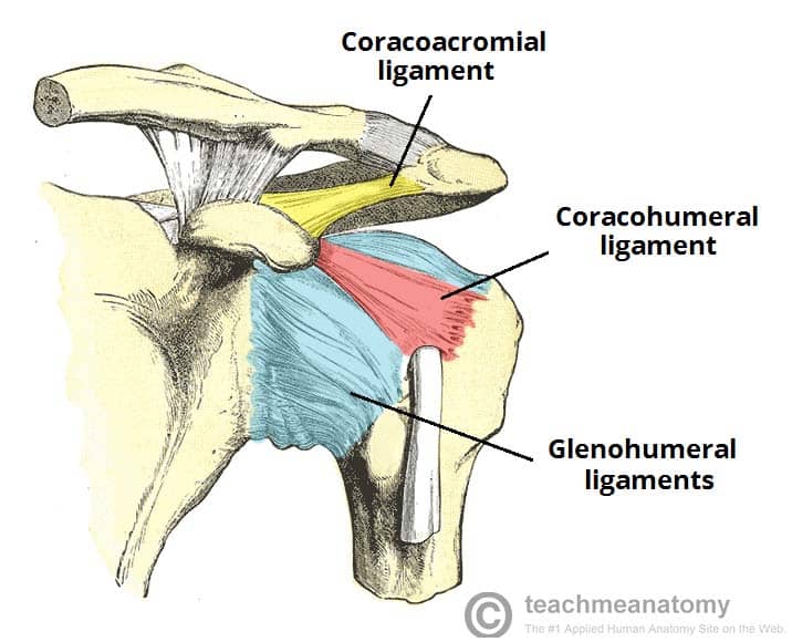
The Shoulder Joint Structure Movement Teachmeanatomy from teachmeanatomy.info The humerus or one of the other bones in the shoulder slips out of position. Around the shoulder, muscles in the back, neck, shoulder, chest and upper arm all work together to support and move the shoulder. Browse 3,107 anatomy of neck and shoulder stock photos and images available, or start a new search to explore more stock photos and images. Cervical nerves are spinal nerves that arise from the cervical region of the spinal cord. Diagram of bones in neck and shoulder. Place your thumbs under the neck and run the inside of your index finger down the length of it. The neck muscles, including the sternocleidomastoid and the trapezius, are responsible for the gross motor movement in the muscular system of the head and neck. We are pleased to provide you with the picture named neck nerves and innervation of shoulder, arm, and hand.
In this episode of eorthopodtv, orthopaedic surgeon randale c.
Working in pairs on the left and right sides of the body, these muscles. The shoulder is one of the largest and most complex joints in the body. The neck is one of the most complex and intricate structures in our body and includes the spinal cord, which sends messages from the brain to the rest of the body. Human body muscles head muscles muscles of the neck neck and shoulder muscles shoulder joint lower back anatomy upper limb anatomy shoulder muscle anatomy human muscle anatomy. We are pleased to provide you with the picture named neck nerves and innervation of shoulder, arm, and hand. The majority of these nerves control the functions of the upper extremities and allow you to feel your arms, shoulder, and back of your head. The clavicle is the only bony attachment between the trunk and the upper limb. We think this is the most useful anatomy picture that you need. They move the head in every direction, pulling the skull and jaw towards the shoulders, spine, and scapula. The shoulder joint is the junction between the chest and the upper extremity. Contains glands ( thyroid, parathyroid, and thymus ), the larynx, pharynx and trachea. You can use your middle, ring, and pinky fingers on the front of the shoulders. Related posts of diagram of the neck anatomy veins and arteries of the neck.
🤔 the acetabulofemoral joint , commonly called the hip joint , scientifically termed is located in between the pelvis and the femur of the legs. • the shoulder muscles may be divided functionally into two groups. The cervical spine is responsible for several crucial roles, including. Contain the common carotid artery, internal. The majority of these nerves control the functions of the upper extremities and allow you to feel your arms, shoulder, and back of your head.
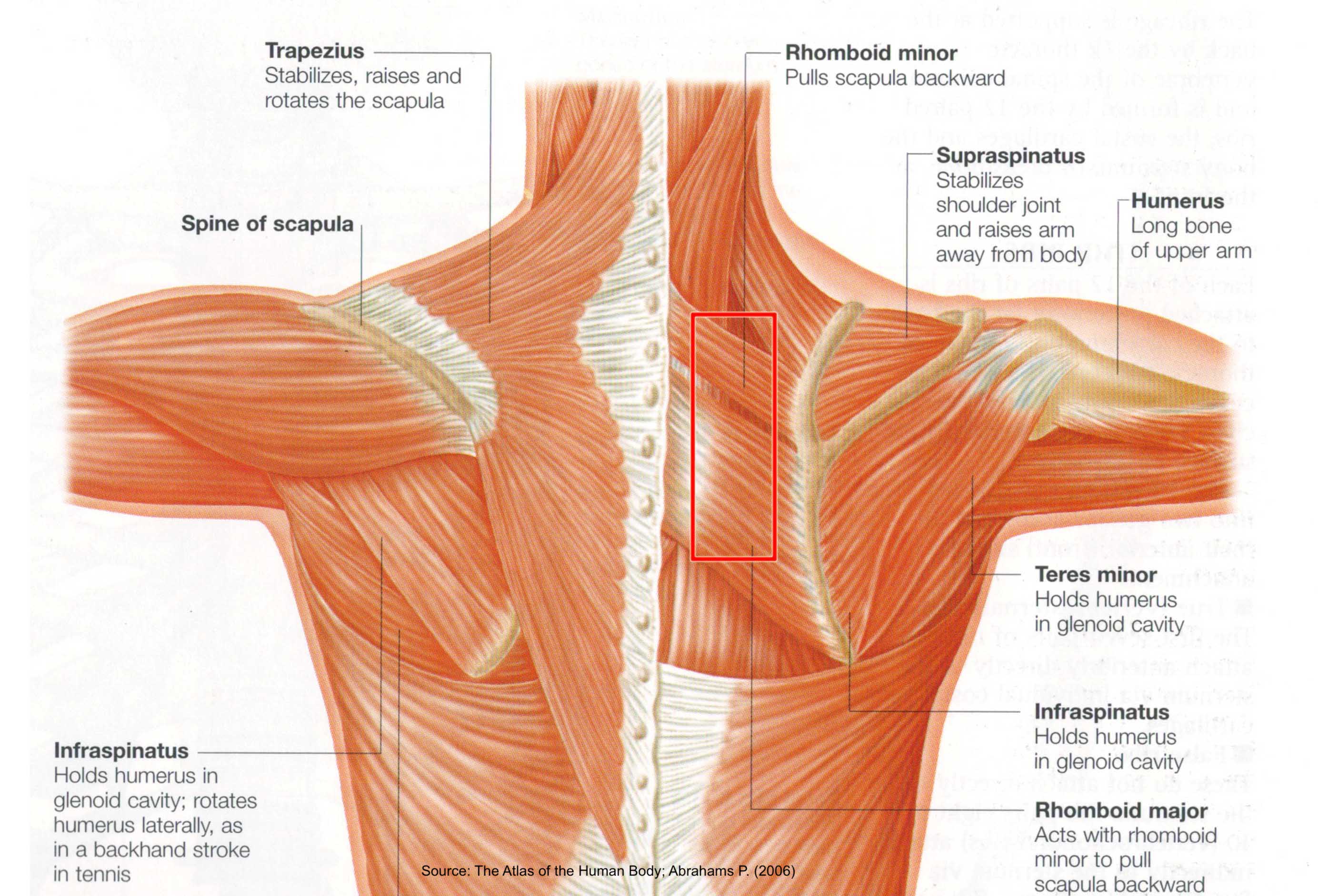
Beautiful Illustration Of The Deep And Superficial Musculature Of The Back This Anatomical Slide By Nett Shoulder Muscle Anatomy Muscle Diagram Muscle Anatomy from i.pinimg.com Fan the motion out over their shoulders as well. Rectus capitis posterior major, which arises from the spinous process of the axis (c2). Contain the common carotid artery, internal. Human body muscles head muscles muscles of the neck neck and shoulder muscles shoulder joint lower back anatomy upper limb anatomy shoulder muscle anatomy human muscle anatomy. 🤔 the acetabulofemoral joint , commonly called the hip joint , scientifically termed is located in between the pelvis and the femur of the legs. The cervical spine is responsible for several crucial roles, including. Begin at the ear and work down to where the neck meets the shoulder. There are eight pairs of cervical nerves, denoted c1 to c8.
Sechrest, md narrates an animated tutorial on the basic anatomy of the shoulder.
The shoulder is one of the largest and most complex joints in the body. The neck is the start of the spinal column and spinal cord. Each nerve provides sensation to a specific area of the body called a dermatome. The majority of these nerves control the functions of the upper extremities and allow you to feel your arms, shoulder, and back of your head. The head and neck is covered in skin and its appendages, termed the integumentary system.these include hair, sweat glands, sebaceous glands, and sensory nerves.the skin is made up of three microscopic layers: A second joint in the shoulder is the junction of the collar bone with the shoulder blade, called the. Cervical nerves are spinal nerves that arise from the cervical region of the spinal cord. Fan the motion out over their shoulders as well. Sechrest, md narrates an animated tutorial on the basic anatomy of the shoulder. The content of the neck is grouped into 4 neck spaces, called the compartments. Bones have many shapes and sizes and are important to add structure to the body and protection to the the shoulder girdle combines to give you shoulder motion. The shoulder is one of the largest and most complex joints in the body. Related posts of diagram of the neck anatomy veins and arteries of the neck.
You can use your middle, ring, and pinky fingers on the front of the shoulders neck anatomy diagram. Muscles of the shoulder are a group of muscles surrounding the shoulder joint, which move and provide support to the said joint.
